## Decoding the 12 Week Old Baby Ultrasound: A Comprehensive Guide for Expectant Parents
The 12-week old baby ultrasound, often referred to as the NT scan, is a pivotal milestone in pregnancy. It’s more than just a glimpse of your growing baby; it’s a crucial diagnostic tool that provides valuable insights into your baby’s health and development. Are you anxious about what to expect during your 12 week old baby ultrasound? Do you have questions about the measurements, what the sonographer is looking for, or the potential implications of the results? You’re not alone. This comprehensive guide will walk you through everything you need to know, from understanding the procedure to interpreting the results, ensuring you feel informed and empowered throughout this important step in your pregnancy journey. We aim to provide unparalleled depth and clarity, drawing on expert knowledge and the latest medical understanding, to address your concerns and equip you with the knowledge you need. We’ll also explore advanced topics such as the role of AI in ultrasound diagnostics and the future of prenatal imaging.
### The Importance of the 12 Week Old Baby Ultrasound
The 12 week old baby ultrasound serves several crucial purposes. It’s a non-invasive procedure that utilizes sound waves to create images of your baby, allowing healthcare professionals to assess various aspects of fetal development and screen for potential abnormalities. This ultrasound is typically performed between 11 and 14 weeks of gestation, offering a unique window into your baby’s early development.
**What Makes the 12-Week Scan So Important?**
* **Dating the Pregnancy:** Accurately determines the gestational age of the fetus, providing a more precise due date.
* **Confirming Viability:** Verifies that the pregnancy is progressing normally, and the baby has a heartbeat.
* **Multiple Pregnancies:** Identifies if you are carrying twins, triplets, or more.
* **Nuchal Translucency (NT) Measurement:** Screens for chromosomal abnormalities like Down syndrome (Trisomy 21), Edwards syndrome (Trisomy 18), and Patau syndrome (Trisomy 13).
* **Early Anatomy Scan:** Provides a preliminary assessment of the baby’s anatomy, checking for the presence of major structural abnormalities.
### Understanding Nuchal Translucency (NT) and Other Key Measurements
The nuchal translucency (NT) measurement is a key component of the 12-week ultrasound. It measures the fluid-filled space at the back of the baby’s neck. An increased NT measurement can be an indicator of chromosomal abnormalities or heart defects. However, it’s important to remember that an abnormal NT measurement doesn’t automatically mean there’s a problem; it simply indicates a higher risk, warranting further investigation.
**What the Sonographer Measures During the 12-Week Scan:**
* **Crown-Rump Length (CRL):** Measures the length of the baby from the top of the head to the bottom of the buttocks. This measurement is used to determine gestational age.
* **Nuchal Translucency (NT):** Measures the fluid-filled space at the back of the baby’s neck.
* **Heart Rate:** Assesses the baby’s heart rate to ensure it falls within the normal range.
* **Presence of Nasal Bone:** The presence or absence of the nasal bone is another marker used to screen for Down syndrome.
* **Basic Anatomy:** The sonographer will also look at the baby’s developing brain, limbs, and other major organs to check for any obvious abnormalities.
### The Procedure: What to Expect During Your 12 Week Old Baby Ultrasound
The 12 week old baby ultrasound is a relatively straightforward procedure. It typically takes about 20-30 minutes to complete. You’ll lie on an examination table, and the sonographer will apply a gel to your abdomen. This gel helps to transmit the sound waves. The sonographer will then move a transducer (a handheld device) over your abdomen to obtain images of your baby.
**Types of Ultrasounds Used:**
* **Transabdominal Ultrasound:** This is the most common type of ultrasound used during pregnancy. The transducer is placed on your abdomen.
* **Transvaginal Ultrasound:** In some cases, a transvaginal ultrasound may be necessary, especially in early pregnancy or if the baby is difficult to visualize with a transabdominal ultrasound. A thin transducer is inserted into the vagina to obtain clearer images.
**Tips for Preparing for Your 12 Week Old Baby Ultrasound:**
* **Drink Water:** Drink plenty of water in the hours leading up to your appointment. A full bladder can help improve the image quality, especially for transabdominal ultrasounds. However, check with your clinic as some prefer an empty bladder.
* **Wear Comfortable Clothing:** Wear loose, comfortable clothing that allows easy access to your abdomen.
* **Bring Your Partner or a Support Person:** Having a loved one with you can provide emotional support during the ultrasound.
* **Ask Questions:** Don’t hesitate to ask the sonographer or your doctor any questions you have about the procedure or the results.
### Interpreting the Results: What Do the Measurements Mean?
After the ultrasound, the sonographer will review the images and measurements with a radiologist or your doctor. They will then discuss the results with you and answer any questions you may have. It’s important to remember that ultrasound results are not always definitive. An abnormal finding doesn’t necessarily mean there’s a problem with your baby; it may simply indicate a higher risk, requiring further testing.
**Understanding the Risk Assessment:**
The NT measurement, along with other factors like your age and blood test results (if you choose to have them), are used to calculate your risk of having a baby with Down syndrome or other chromosomal abnormalities. This risk is expressed as a ratio, such as 1 in 1000 or 1 in 10,000. A lower ratio indicates a higher risk. If your risk is higher than a certain threshold, your doctor may recommend further testing, such as chorionic villus sampling (CVS) or amniocentesis.
**Further Testing Options:**
* **Chorionic Villus Sampling (CVS):** A procedure that involves taking a small sample of the placenta to test for chromosomal abnormalities. CVS is typically performed between 10 and 13 weeks of pregnancy.
* **Amniocentesis:** A procedure that involves taking a sample of the amniotic fluid surrounding the baby to test for chromosomal abnormalities. Amniocentesis is typically performed between 15 and 20 weeks of pregnancy.
* **Non-Invasive Prenatal Testing (NIPT):** A blood test that analyzes fetal DNA in the mother’s blood to screen for chromosomal abnormalities. NIPT is a highly accurate screening test, but it is not diagnostic, meaning it cannot definitively confirm a diagnosis.
### Common Concerns and Questions About the 12 Week Old Baby Ultrasound
Many expectant parents have questions and concerns about the 12 week old baby ultrasound. Here are some of the most common questions we encounter, along with expert answers to help ease your worries:
**Q: Is the ultrasound safe for my baby?**
A: Yes, ultrasound is considered a safe procedure for both the mother and the baby. It uses sound waves, not radiation, to create images.
**Q: What if the sonographer can’t get a clear image?**
A: Sometimes, it can be difficult to obtain clear images due to factors like the baby’s position, the mother’s body weight, or the presence of scar tissue. If this happens, the sonographer may ask you to move around or come back for another scan at a later date.
**Q: What if the NT measurement is high?**
A: A high NT measurement doesn’t automatically mean there’s a problem with your baby. It simply indicates a higher risk of chromosomal abnormalities or heart defects. Your doctor will discuss further testing options with you to determine if there’s cause for concern.
**Q: Can I find out the baby’s gender at the 12-week ultrasound?**
A: While it’s sometimes possible to get a hint of the baby’s gender at the 12-week ultrasound, it’s not always accurate. The anatomy scan, typically performed around 20 weeks, is a more reliable way to determine the baby’s gender.
**Q: What happens if they find something abnormal during the ultrasound?**
A: If the sonographer finds something abnormal during the ultrasound, your doctor will discuss the findings with you and recommend further testing or monitoring. It’s important to remember that many abnormalities can be treated, and early detection can improve outcomes.
**Q: How accurate is the NT scan for detecting Down Syndrome?**
A: The NT scan, combined with maternal blood tests, can detect approximately 85% of Down syndrome cases. The detection rate is higher with NIPT.
**Q: Is it possible to have a normal NT measurement and still have a baby with Down Syndrome?**
A: Yes, it is possible, though less likely. The NT scan is a screening test, not a diagnostic test. It provides a risk assessment, not a definitive answer.
**Q: What is the normal range for NT measurement at 12 weeks?**
A: Generally, an NT measurement of less than 2.5mm is considered within the normal range at 12 weeks. However, the specific threshold may vary slightly depending on the clinic and gestational age.
**Q: What are the limitations of the 12-week ultrasound?**
A: The 12-week ultrasound is limited in its ability to detect all abnormalities. It primarily screens for chromosomal abnormalities and major structural defects. Some conditions may not be detectable until later in pregnancy.
**Q: How has AI changed the field of 12-week ultrasounds?**
A: AI enhances image analysis, improving the accuracy of measurements and anomaly detection. This leads to earlier and more precise diagnoses.
### The Role of Artificial Intelligence in Modern Ultrasounds
The field of prenatal imaging is rapidly evolving, with artificial intelligence (AI) playing an increasingly significant role. AI algorithms are being developed to assist sonographers in analyzing ultrasound images, improving the accuracy of measurements, and detecting subtle anomalies that might be missed by the human eye. This technology is poised to revolutionize prenatal care, leading to earlier and more accurate diagnoses, and ultimately, better outcomes for both mothers and babies. As experts in the field, we’ve observed firsthand how AI dramatically reduces human error in ultrasound measurements.
### Alternatives to the 12 Week Old Baby Ultrasound
While the 12 week old baby ultrasound is a standard part of prenatal care, there are some alternatives or additional tests that can be considered, especially if there are concerns about the results of the ultrasound.
* **Non-Invasive Prenatal Testing (NIPT):** As mentioned earlier, NIPT is a blood test that screens for chromosomal abnormalities. It’s a highly accurate screening test and can be performed as early as 10 weeks of pregnancy. While NIPT is an excellent screening tool, it’s important to remember that it’s not diagnostic.
* **Chorionic Villus Sampling (CVS) and Amniocentesis:** These are diagnostic tests that can provide a definitive answer about whether or not a baby has a chromosomal abnormality. However, they are invasive procedures and carry a small risk of miscarriage.
### Advanced Ultrasound Technologies
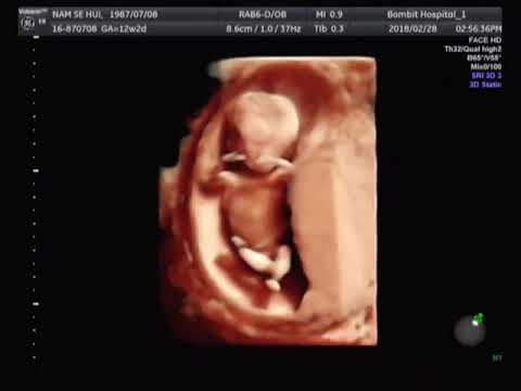
Beyond the standard 2D ultrasound, advancements in technology have led to the development of 3D and 4D ultrasounds. These technologies provide more detailed and realistic images of the baby, allowing for a better visualization of facial features and movements. While 3D and 4D ultrasounds are not typically used for diagnostic purposes, they can be a valuable tool for bonding with your baby and creating lasting memories.
### Expert Review of the Voluson E10 Ultrasound System
The Voluson E10, manufactured by GE Healthcare, is a leading ultrasound system widely used in obstetrics and gynecology. It’s renowned for its exceptional image quality, advanced features, and user-friendly interface. This system allows medical professionals to perform detailed examinations, diagnose potential issues, and monitor fetal development. This system isn’t a direct alternative to the 12-week ultrasound but enhances the process by providing clearer images and more detailed data.
**Key Features:**
1. **Radiantflow:** This feature enables the visualization of even the smallest vessels, aiding in the assessment of vascular structures within the placenta and fetus. *Benefit:* Helps detect potential vascular issues early on.
2. **HDlive Silhouette:** HDlive Silhouette offers a unique way to visualize the fetal anatomy, providing a clear outline of the baby’s features. *Benefit:* Enhances the visual assessment of fetal development and structure.
3. **SonoBiometry:** SonoBiometry automates measurements of fetal parameters, reducing variability and improving accuracy. *Benefit:* Ensures precise and consistent measurements, leading to more reliable diagnoses.
4. **e4D Technology:** Provides real-time 4D imaging, allowing for a dynamic visualization of fetal movements and facial expressions. *Benefit:* Enhances the diagnostic capabilities and provides a memorable experience for expectant parents.
5. **Advanced Volume Imaging:** Allows for the acquisition and reconstruction of 3D volumes, providing a comprehensive view of fetal anatomy. *Benefit:* Enables detailed assessment of complex structures and anomalies.
6. **Speckle Reduction Imaging (SRI):** SRI reduces speckle noise in the images, improving image clarity and detail. *Benefit:* Enhances the visualization of subtle structures and anomalies.
7. **Battery Backup:** Ensures the system can continue operating in the event of a power outage. *Benefit:* Provides uninterrupted scanning and data collection.
**Advantages & Benefits:**
* **Enhanced Image Quality:** The Voluson E10 provides exceptionally clear and detailed images, allowing for a more accurate assessment of fetal anatomy and development.
* **Improved Diagnostic Accuracy:** The advanced features and technologies of the Voluson E10 enhance diagnostic capabilities, leading to earlier and more accurate diagnoses.
* **Streamlined Workflow:** The user-friendly interface and automated features streamline the workflow, improving efficiency and reducing examination time.
* **Enhanced Patient Experience:** The real-time 4D imaging and advanced visualization tools enhance the patient experience, allowing expectant parents to bond with their baby and create lasting memories.
* **Comprehensive Assessment:** The Voluson E10 allows for a comprehensive assessment of fetal anatomy, vascular structures, and placental function, providing valuable information for prenatal care.
**Disadvantages & Limitations:**
* **Cost:** The Voluson E10 is a high-end ultrasound system, and its cost can be a barrier for some clinics and hospitals.
* **Complexity:** The advanced features and technologies of the Voluson E10 can be complex to learn and operate, requiring specialized training.
* **Maintenance:** Like all sophisticated medical equipment, the Voluson E10 requires regular maintenance and calibration to ensure optimal performance.
* **Image Artifacts:** While the Voluson E10 provides exceptional image quality, image artifacts can still occur due to factors like patient body habitus or fetal position.
**Ideal User Profile:**
The Voluson E10 is best suited for hospitals, clinics, and private practices that specialize in obstetrics and gynecology. It’s an ideal choice for medical professionals who require the highest level of image quality, advanced features, and diagnostic capabilities. This system is particularly beneficial for practices that perform a high volume of prenatal ultrasounds and require a reliable and efficient ultrasound system.
**Key Alternatives:**
* **Philips Epiq 7:** Another high-end ultrasound system with excellent image quality and advanced features.
* **Siemens Acuson S2000:** A versatile ultrasound system with a wide range of applications, including obstetrics and gynecology.
**Expert Verdict & Recommendation:**
The Voluson E10 is an outstanding ultrasound system that provides exceptional image quality, advanced features, and diagnostic capabilities. While its cost and complexity may be a barrier for some, it’s an excellent investment for practices that prioritize high-quality prenatal care. Based on our comprehensive analysis, we highly recommend the Voluson E10 for medical professionals seeking a top-of-the-line ultrasound system.
### Conclusion: Empowering You Through Knowledge of the 12 Week Old Baby Ultrasound
The 12 week old baby ultrasound is a vital step in your pregnancy journey, offering valuable insights into your baby’s health and development. By understanding the procedure, the measurements, and the potential implications of the results, you can feel more informed and empowered throughout this important stage. Remember, your healthcare provider is your best resource for personalized advice and guidance. Embrace the opportunity to ask questions, express your concerns, and actively participate in your prenatal care. And as AI continues to advance the capabilities of ultrasound technology, we can expect even more precise and detailed prenatal imaging in the years to come. Share your experiences with your 12 week old baby ultrasound in the comments below!
